Fungal Skin Discoloration Quiz
This quiz helps determine if your skin symptoms could be related to fungal discoloration or other skin conditions.
Result:
Quick Take
- Fungal skin discoloration is usually caused by Malassezia overgrowth, showing up as patches of lighter or darker skin.
- It often appears on the chest, back, and shoulders where sweat and oil accumulate.
- The condition can masquerade as eczema, psoriasis, or vitiligo, leading to misdiagnosis.
- Topical azoles and short courses of oral antifungals clear most cases within weeks.
- Persistent discoloration may need pigment‑restoring therapies or a dermatologist’s evaluation.
What is fungal skin discoloration?
When most people hear "fungal infection" they picture a red, itchy rash. Tinea versicolor is a common fungal skin disorder that causes patches of altered color-either lighter (hypopigmentation) or darker (hyperpigmentation) than the surrounding skin. It’s not an aggressive infection; the fungus lives quietly on the skin’s surface, waiting for the right conditions to multiply.
The culprit is a yeast‑like fungus called Malassezia a lipophilic yeast that thrives on sebum and sweat. Under normal circumstances, Malassezia co‑exists peacefully with other skin microbes. When humidity, heat, or a weakened immune response tip the balance, the yeast proliferates, producing acids that interfere with normal melanin production. The result? Those blotchy spots that seem to appear overnight.
How does the condition develop?
Three main factors set the stage for a flare:
- Excess oil and sweat: Warm climates, intense workouts, or tight clothing trap moisture, giving Malassezia a buffet.
- UV exposure: Sunlight temporarily masks the discoloration, making the patches look normal. As the skin tans, the affected areas stay their original shade, creating a stark contrast.
- Immune shifts: Illness, hormonal changes, or certain medications can dampen the skin’s natural defenses, allowing the yeast to overgrow.
Because the fungus lives on the surface, it’s easy to treat once identified-provided the diagnosis is spot‑on.
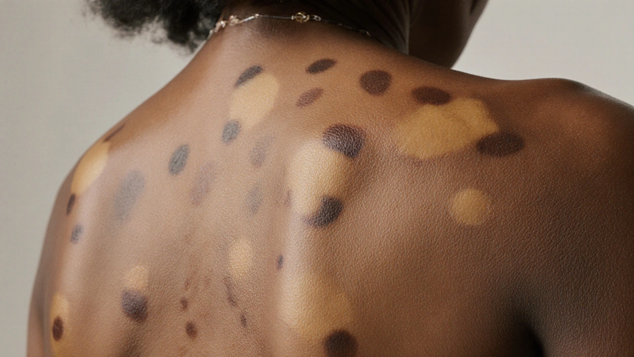
Visual clues: hyperpigmentation vs hypopigmentation
People often mistake the pattern for other disorders. Here’s what to look for:
- Hypopigmented patches: Light‑colored, often pink‑ish, with a fine scaling that’s more noticeable after a hot shower.
- Hyperpigmented patches: Darker brown spots, usually on the upper back or chest, with a subtle, powdery feel.
- Distribution: The rash respects natural skin creases-think neck, upper torso, and sometimes the arms.
If the spots worsen after sun exposure or fade in the shade, fungal discoloration is a strong suspect.

Overlap with other skin disorders
Fungal discoloration shares several visual traits with more chronic conditions. Understanding the differences can save months of frustration.
| Condition | Primary Cause | Typical Appearance | Usual Locations | First‑line Treatment |
|---|---|---|---|---|
| Tinea versicolor | Malassezia overgrowth | Light or dark patches with fine scaling | Chest, back, shoulders | Topical azoles or oral itraconazole |
| Eczema (Atopic Dermatitis) | Genetic & environmental triggers | Red, itchy patches, sometimes weepy | Inner elbows, behind knees, face | Moisturizers, topical steroids, calcineurin inhibitors |
| Psoriasis | Autoimmune inflammation | Silvery‑scale plaques | Scalp, elbows, knees, lower back | Topical vitamin D analogs, biologics |
| Vitiligo | Autoimmune loss of melanocytes | Well‑defined white patches | Anywhere, often around orifices | Topical steroids, phototherapy |
Notice how the cause, texture, and treatment differ. The table helps clinicians and readers quickly rule in or out fungal discoloration.
Why misdiagnosis happens
Even seasoned dermatologists can be tripped up. The three biggest reasons:
- Similar color changes: Both vitiligo and tinea versicolor can produce light patches, but vitiligo lacks scaling and won’t bounce back after a warm shower.
- Shared itching: Eczema and fungal discoloration may itch, yet eczema’s rash is usually more inflamed and responsive to steroids.
- Sun‑masking effect: UV light temporarily hides the contrast, making the fungal spots look like “normal” skin until the sun fades.
A simple KOH (potassium hydroxide) skin scrape under a microscope reveals the characteristic “spaghetti‑and‑meatball” appearance of Malassezia-an easy test that clinches the diagnosis.
Managing fungal skin discoloration
Once confirmed, treatment is straightforward. Here’s a roadmap most patients follow:
- Topical antifungals: Creams or shampoos containing selene‑d (selenium sulfide), ketoconazole, or ciclopirox are first‑line. Apply once or twice daily for 2‑4 weeks.
- Oral antifungals: For extensive or recurrent cases, a short course (1-2 weeks) of fluconazole or itraconazole can clear the yeast from deeper skin layers.
- Adjunct skin‑lightening agents: If discoloration lingers, products with azelaic acid or niacinamide can help even out tone.
- Lifestyle tweaks: Keep skin dry, wear breathable fabrics, and limit excessive sun exposure. Shower promptly after sweating.
Most people see a visible improvement within a week, but full color normalization may take a month or two.
When to suspect another disorder
If any of these red flags appear, it’s time to broaden the differential diagnosis:
- Rapid expansion of patches beyond the typical torso area.
- Severe itching, burning, or oozing that doesn’t respond to antifungals.
- Presence of well‑defined, sharply bordered white spots (think vitiligo).
- Scaly plaques with a silvery sheen (classic psoriasis sign).
In such cases, a dermatologist may order a skin biopsy or specific blood tests to rule out autoimmune involvement.
Patient checklist: Spot‑check your skin
- Do the patches change color after a hot shower?
- Are they on oily areas like the upper back or chest?
- Is there a fine, powdery scaling?
- Do the spots become more noticeable after sun exposure?
- Have antifungal creams helped at all?
If you answered “yes” to most of these, fungal skin discoloration is likely. Still, a quick visit to a healthcare professional can confirm it and prevent unnecessary treatments.
Frequently Asked Questions
Can tinea versicolor spread to other parts of the body?
Yes, the yeast can colonize any oily skin region. However, it usually stays on the torso, shoulders, and upper arms because those areas produce the most sebum.
Why does my skin look normal in the shade but darker in the sun?
Sunlight triggers melanin production in healthy skin, creating a tan. The fungus‑affected zones can’t ramp up melanin, so they stay their original shade, making the contrast obvious.
Are over‑the‑counter shampoos effective?
Shampoos with selenium sulfide or zinc pyrithione work well for mild cases because they coat the skin and reduce yeast load. For stubborn patches, a prescription cream is often needed.
Can I get tinea versicolor from my pet?
No. Malassezia lives naturally on human skin; pets carry different fungal species. Transmission between people is rare and usually linked to shared hot, humid environments.
Will the discoloration ever come back?
Recurrence is common, especially in warm climates or after heavy sweating. Maintenance with a weekly anti‑fungal wash can keep the yeast in check.


Stephen Jahl
September 29, 2025 AT 22:28In the ontological framework of dermatological pathophysiology, the over-proliferation of Malassezia spp. constitutes a quintessential example of dysbiosis mediated melanogenesis perturbation. The phenomenology elucidated herein, wherein sebum-rich microenvironments catalyze ergosterol synthesis, aligns with the canonical paradigm of lipid-dependent mycotic colonization. Consequently, therapeutic regimens predicated upon azole inhibition of lanosterol 14α‑demethylase present a mechanistic rationale of considerable clinical pertinence. One must, however, remain circumspect towards iatrogenic resistance patterns emergent from subtherapeutic dosing.
gershwin mkhatshwa
October 3, 2025 AT 04:28Yo, this stuff is crazy how a little yeast can mess up your skin tone. If you’re hanging at the gym a lot, make sure you shower right after, otherwise you’re just feeding the fungus. Keep an eye on the scaling after a hot shower – that’s the tell.
Samantha Kolkowski
October 6, 2025 AT 10:28Hey there! i think the table you posted is super helpful, just remember to keep the skin dry after workouts.
Nick Ham
October 9, 2025 AT 16:28Malassezia overgrowth is a lipid‑dependent mycotic dysbiosis; azole therapy targets ergosterol biosynthesis.
Jennifer Grant
October 12, 2025 AT 22:28When you consider the broader sociocultural implications of skin disorders, it becomes evident that fungal discoloration is not merely a superficial annoyance but a reflection of how modern lifestyles intersect with microbial ecology. The relentless pursuit of high‑intensity fitness regimes, coupled with the ubiquitous use of synthetic fabrics, creates a perfect micro‑climate for Malassezia to flourish. Moreover, the psychological burden of visible skin changes can erode self‑esteem, leading to a cascade of social withdrawal that is often overlooked in clinical discussions. Historically, dermatology has privileged hyperpigmentation disorders such as melasma, relegating tinea versicolor to a footnote, despite its prevalence across diverse populations. In the diagnostic algorithm, a simple potassium hydroxide (KOH) preparation can reveal the classic spaghetti‑and‑meatball morphology, yet many primary care providers bypass this step in favor of empiric steroid prescriptions. This practice not only delays appropriate antifungal therapy but also risks exacerbating the fungal load by suppressing local immune responses. From a therapeutic standpoint, topical azoles like ketoconazole have a well‑documented safety profile, but adherence falters when patients perceive the rash as cosmetic rather than pathological. Oral agents such as itraconazole provide a systemic sweep, but clinicians must weigh hepatic considerations, especially in patients with comorbidities. Lifestyle modifications, though often dismissed as trivial, play a pivotal role; breathable clothing, prompt post‑exercise cleansing, and judicious sun exposure can tip the balance back toward homeostasis. The interplay between ultraviolet radiation and melanin synthesis further complicates the clinical picture, as UV can mask hypopigmented patches, only to reveal stark contrast once tanning subsides. Public health messaging, therefore, should emphasize not just treatment, but prevention through environmental control. In communities with high humidity, community‑wide education on skin hygiene can reduce outbreak clusters. Finally, the psychosocial dimension warrants integrated care, perhaps involving counselors to address body image concerns that linger even after the fungus is eradicated. In sum, the management of fungal skin discoloration is a multifaceted endeavor that demands both biomedical precision and empathetic patient engagement. Thus, a collaborative approach between dermatologist, primary care, and patient is essential for lasting resolution.
Kenneth Mendez
October 16, 2025 AT 04:28They dont want you to know that the pharma companies push those creams just to keep us buying more.
Gabe Crisp
October 19, 2025 AT 10:28The moral imperative is to seek evidence‑based treatments rather than succumb to sensational narratives.
Paul Bedrule
October 22, 2025 AT 16:28The epistemic contours of cutaneous mycology demand a dialectic between empirical observation and phenomenological interpretation, lest we reduce complex dermal symbiosis to mere pathologization.
yash Soni
October 25, 2025 AT 22:28So basically, wash your back and stop overthinking it.
Emily Jozefowicz
October 29, 2025 AT 03:28Oh great, another “miracle cure” that’s just a fancy shampoo. If only we could bottle the enthusiasm of dermatologists in a bottle of anti‑fungal shampoo, we’d all be flawless.
Franklin Romanowski
November 1, 2025 AT 09:28While humor lightens the mood, remember that skin health is a holistic journey; combining proper hygiene with compassionate self‑care cultivates both confidence and clarity.
Brett Coombs
November 4, 2025 AT 15:28Honestly, I think the whole “sun masks the patches” thing is just an excuse for people to blame the weather instead of their own laziness.
John Hoffmann
November 7, 2025 AT 21:28The statement oversimplifies a complex photobiological interaction; UV exposure does not merely “mask” but alters melanogenic pathways, yielding differential pigmentation.
Shane matthews
November 11, 2025 AT 03:28Just a reminder keep skin dry and use a gentle anti fungal cream it works for most cases
Rushikesh Mhetre
November 14, 2025 AT 09:28Exactly!!! Make sure to shower right after a sweaty workout, dry thoroughly, and apply the antifungal-twice daily if needed!!! Consistency is the key!!! 💪
Sharath Babu Srinivas
November 17, 2025 AT 15:28Indeed, adherence to the regimen maximizes efficacy; moreover, intermittent use can lead to recurrence 😐👍.
Gauri Omar
November 20, 2025 AT 21:28Listen up: if you keep ignoring the warning signs, the fungus will come back stronger, and nothing will stop it but a disciplined, relentless routine that respects your skin’s sovereignty.